When starting orthodontic treatment, diagnostic imaging plays a critical role in building an effective treatment plan. If you’ve researched braces or Invisalign, you may have come across two types of imaging: traditional 2D X-rays and CBCT scans.
Both technologies help orthodontists evaluate teeth, roots, jaw alignment, and facial structure—but they are not the same. Understanding the differences can help you feel more confident about your treatment decisions.
What Are 2D Dental X-Rays?
Two-dimensional (2D) X-rays have been used in dentistry and orthodontics for decades. These images provide flat representations of your teeth and jaw structures.
Common types include:
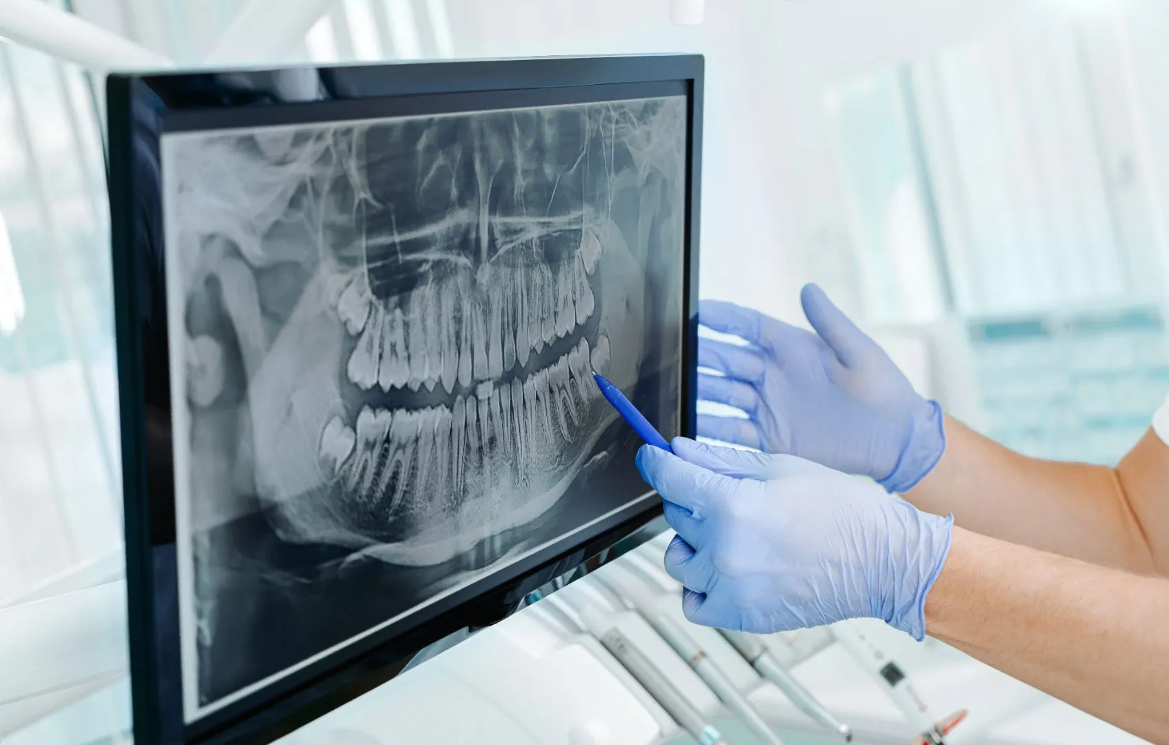
- Panoramic X-rays — Show all teeth, upper and lower jaws, and surrounding structures in a single image
- Cephalometric X-rays — Capture a side view of the head to analyze jaw growth and facial alignment
- Bitewing X-rays — Focus on specific areas to detect decay or bone levels
2D imaging is fast, widely available, and exposes patients to relatively low radiation levels. For many straightforward orthodontic cases, it provides enough information to plan treatment effectively.
However, because these images are flat, they do not show depth. Structures can overlap, and certain details may not be fully visible.
What Is CBCT Imaging?
CBCT stands for Cone Beam Computed Tomography. Unlike traditional X-rays, CBCT produces three-dimensional (3D) images of the teeth, jaws, roots, and surrounding structures.
Instead of a flat picture, CBCT creates a detailed digital model that can be viewed from multiple angles. This allows orthodontists to evaluate:
- Tooth root positioning
- Bone density and thickness
- Impacted or unerupted teeth
- Airway space
- Jaw joint structure (TMJ)
- Relationship between nerves and teeth
CBCT provides a more comprehensive view of the entire craniofacial structure, which can be especially helpful in complex cases.
Accuracy: 2D vs. 3D Imaging
When comparing CBCT vs. 2D X-rays for braces, accuracy largely depends on the complexity of the case.
Situations Where 2D Imaging May Be Sufficient
For mild to moderate crowding or spacing issues, traditional 2D X-rays often provide enough information to create an effective treatment plan. These images can clearly show tooth positioning, general jaw relationships, and root length.
In many routine orthodontic cases, 2D imaging remains a reliable and efficient diagnostic tool.
Situations Where CBCT May Offer Greater Accuracy
CBCT imaging can provide greater diagnostic precision in cases involving:
- Impacted teeth — Especially canines that are trapped beneath the gum line
- Severe bite discrepancies — Such as significant underbites or asymmetry
- Jaw growth abnormalities — Where 3D evaluation improves treatment planning
- TMJ concerns — When evaluating joint structure and alignment
- Airway analysis — Particularly in patients with breathing concerns
Because CBCT shows depth and spatial relationships, it reduces guesswork and can reveal details that may not be visible on flat images.
Radiation Exposure: Is One Safer?
Radiation exposure is an important consideration for many families.
Traditional 2D X-rays generally expose patients to lower radiation compared to CBCT scans. However, modern CBCT technology has significantly reduced radiation levels compared to earlier versions and medical CT scans.
Orthodontists follow strict guidelines when determining whether CBCT imaging is necessary. The goal is always to use the lowest radiation dose possible while still obtaining the information needed for safe and effective treatment.
CBCT is typically reserved for cases where the additional detail provides meaningful clinical value.
Treatment Planning Differences
The level of imaging detail can influence how braces or Invisalign treatment is planned.
With 2D imaging, orthodontists assess alignment, spacing, and jaw relationships using flat views. For many patients, this provides sufficient guidance for predictable outcomes.
With CBCT imaging, orthodontists can:
- Visualize exact root angulation
- Assess bone support before moving teeth
- Identify hidden anatomical variations
- Plan surgical or complex orthodontic procedures more precisely
This level of insight can be particularly helpful when treatment involves impacted teeth, jaw discrepancies, or multidisciplinary care with oral surgeons.
Is CBCT Always Necessary for Braces?
Not necessarily.
Many orthodontic cases are successfully treated using digital scans combined with traditional 2D X-rays. CBCT is typically recommended only when additional diagnostic clarity is needed.
Imaging decisions are based on individual factors such as:
- Age
- Growth stage
- Complexity of the bite
- Presence of impacted teeth
- Jaw alignment concerns
The goal is to choose the imaging method that provides enough information for safe, effective treatment—without unnecessary exposure.
Benefits of Advanced Imaging in Orthodontics
When used appropriately, advanced imaging technologies can improve outcomes by:
- Increasing diagnostic precision
- Supporting more customized treatment planning
- Reducing unexpected complications
- Improving communication with patients through visual explanations
Three-dimensional imaging also allows patients to better understand their treatment, since they can see a model of their own anatomy from multiple angles.
Which Imaging Is More Accurate?
CBCT imaging provides more detailed and comprehensive information than traditional 2D X-rays. From a purely technical standpoint, 3D imaging offers greater accuracy because it eliminates overlapping structures and provides depth perception.
However, “more accurate” does not always mean “necessary.”
For many patients, traditional 2D imaging provides sufficient diagnostic information to achieve excellent orthodontic results. CBCT becomes particularly valuable when a case involves complexity that cannot be fully evaluated with flat images.
Ultimately, the most appropriate imaging method depends on the individual patient’s needs.
Final Thoughts on CBCT vs. 2D X-Rays
Both CBCT and traditional 2D X-rays play important roles in modern orthodontics. Each has advantages, and both can contribute to safe, effective treatment planning.
If you’re considering braces or Invisalign and have questions about diagnostic imaging, it’s helpful to discuss which option is best for your specific situation.
At Canton Orthodontics, advanced imaging technology is used thoughtfully to ensure precise diagnosis and personalized treatment planning. If you're exploring orthodontic treatment in Canton, GA, schedule a consultation to learn which imaging approach is right for your smile.